

Theragnostic nanocapsules - nuclear medicine
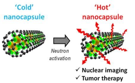
We have developed radioactive nanocapsules that allow nuclear imaging in an ultrasensitive manner and lung cancer therapy. Two different strategies have been employed for the preparation of ‘hot’ radioactive nanocapsules. The first strategy consists in the direct encapsulation of radionuclides in their cavities of the carbon nanocapsules. The second strategy goes via the initial encapsulation of a ‘cold’ isotopically enriched isotope (152Sm), which can then be activated on demand to their ‘hot’ radioactive form (153Sm) by neutron irradiation. The use of ‘cold’ isotopes avoids the need for radioactive facilities during the preparation of the nanocapsules, reduces radiation exposure to personnel, prevents the generation of nuclear waste, and evades the time constraints imposed by the decay of radionuclides. A high specific radioactivity (up to 11.37 GBq/mg) has been achieved by neutron irradiation, making the “hot” nanocapsules useful not only for in vivo imaging but also therapeutically effective against lung cancer metastases after intravenous injection. The external surface remains available and can be funtionalized to increase the dispersability, biocompatibility and for targeting purposes. Glycans, peptides and antibodies have been employed in the performed studies. The high in vivo stability of the radioactive payload, selective toxicity to cancerous tissues, and the elegant preparation method offer a paradigm for application of nanomaterials in radiotherapy.

Neutron capture therapy
Neutron capture therapy (NCT) is a high-linear energy transfer form of radiotherapy that exploits the potential of some specific isotopes for cancer treatment, based on the neutron capture and emission of short-range charged particles, which occur at low energies. The nuclear reaction that takes place when some isotopes are irradiated with low-energy thermal neutrons, produces high linear energy transfer (LET) particles suitable for cancer cell eradication. The limited path lengths of the LET particles (5-9 µm) produced in NCT can limit the destructive effects to isotopes that are localized in cells. Thus, conferring high therapeutic precision to this type of radiotherapy. We are developing several nanocarriers with the aim to increase the amount of neutron capture elements that are delivered to cancer cells (patent application 202230271).
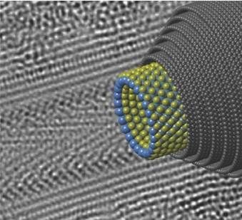
1D tubular van der Waals heterostructures
The electronic and optical properties of two-dimensional layered materials allow the miniaturization of nanoelectronic and optoelectronic devices in a competitive manner. Even larger opportunities arise when two or more layers of different materials are combined. We have observed that the cavities of carbon nanotubes can be employed for the template assisted growth of inorganic metal halide nanotubes in their interior, thus forming 1D tubular van der waals heterostructures. We have developed strategies that result in a high selectivity toward the growth of such 1D heterostructures. A decrease of the resistivity as well as a significant increase in the current flow upon illumination has been observed in a PbI2@CNT bulk matrix. Both effects are attributed to the presence of single-walled lead iodide nanotubes in the cavities of carbon nanotubes (CNTs), which dominate the properties of the whole matrix.


Partnerships for Technology Transfer


Selected Outreach Activities


Selected recent publications
Selected recent publications
Nanomaterials characterization
UV-Vis quantification of the iron content in iteratively steam and HCl purified single-walled carbon nanotubes. Martincic M., Tobías-Rossell G., PLoS ONE. 2024, 19, e0303359. https://doi.org/10.1371/journal.pone.0303359
Thermal Stability and Purity of Graphene and Carbon Nanotubes: Key Parameters for Their Thermogravimetric Analysis (TGA). Martincic M., Sandoval S., Oró-Solé J., & Tobías-Rossell G., Nanomaterials 2024, 14, 1754. https://doi.org/10.3390/nano14211754
Neutron capture therapy
Lithium halide filled carbon nanocapsules: Paving the way towards lithium neutron capture therapy (LiNCT). Gonçalves G., Sandoval S., Llenas M., Balleteros B., Da Ros T., Bortolussi S., Cansolino L., Ferrarie C., Postuma I., Protti N., Melle-Franco M., Altieri S., Tobías-Rossell G., Carbon 2023, 208, 148-159. https://doi.org/10.1016/j.carbon.2023.03.034
Theragnostic nanocapsules
Functionalization of filled radioactive multi-walled carbon nanocapsules by arylation reaction forin vivodelivery of radio-therapy. Gajewska A., Wang J.T., Klippstein R., Martincic M., Pach E., Feldman R., Saccavini J.-C., Tobias G., Ballesteros B., Al-Jamal K.T., Da Ros T., J. Mater. Chem. B. 2022, 10 (1), 47-56. https://doi.org/10.1039/d1tb02195h
Neutron-irradiated antibody-functionalised carbon nanocapsules for targeted cancer radiotherapy. Wang J.T.-W., Spinato C., Klippstein R., Costa P.M., Martincic M., Pach E., Ruiz de Garibay A.P., Ménard-Moyon C., Feldman R., Michel Y., Šefl M., Kyriakou I., Emfietzoglou D., Saccavini J.-C., Ballesteros B., Tobias G., Bianco A., Al-Jamal K.T., Carbon 2020, 162, 410-422. https://doi.org/10.1016/j.carbon.2020.02.060
Neutron Activated 153Sm Sealed in Carbon Nanocapsules for in Vivo Imaging and Tumor Radiotherapy. Wang J.T.-W., Klippstein R., Martincic M., Pach E., Feldman R., Šefl M., Michel Y., Asker D., Sosabowski J.K., Kalbac M., Da Ros T., Ménard-Moyon C., Bianco A., Kyriakou I., Emfietzoglou D., Saccavini J.-C., Ballesteros B., Al-Jamal K.T., Tobias G., ACS Nano 2020, 14 (1) 129-141. https://doi.org/10.1021/acsnano.9b04898
Non-cytotoxic carbon nanocapsules synthesized via one-pot filling and end-closing of multi-walled carbon nanotubes. Martincic M., Vranic S., Pach E., Sandoval S., Ballesteros B., Kostarelos K., Tobias G., Carbon 2019, 141, 782-793 https://doi.org/10.1016/j.carbon.2018.10.006
Evaluation of the immunological profile of antibody-functionalized metal-filled single-walled carbon nanocapsules for targeted radiotherapy. Perez Ruiz De Garibay A., Spinato C., Klippstein R., Bourgognon M., Martincic M., Pach E., Ballesteros B., Ménard-Moyon C., Al-Jamal K.T., Tobias G., Bianco A., Sci. Rep. 2017, 7, 42605 https://doi.org/10.1038/srep42605
Carbon nanotubes allow capture of krypton, barium and lead for multichannel biological X-ray fluorescence imaging. Serpell C.J., Rutte R.N., Geraki K., Pach E., Martincic M., Kierkowicz M., De Munari S., Wals K., Raj R., Ballesteros B., Tobias G., Anthony D.C., Davis B.G., Nat. Commun. 2016, 7, 13118 https://doi.org/10.1038/ncomms13118
Design of antibody-functionalized carbon nanotubes filled with radioactivable metals towards a targeted anticancer therapy. Spinato C., Perez Ruiz De Garibay A., Kierkowicz M., Pach E., Martincic M., Klippstein R., Bourgognon M., Wang J.T.-W., Ménard-Moyon C., Al-Jamal K.T., Ballesteros B., Tobias G., Bianco A., Nanoscale 2016, 8 (25), 12626-12638 https://doi.org/10.1039/c5nr07923c
Filled and glycosylated carbon nanotubes for in vivo radioemitter localization and imaging. Hong S.Y., Tobias G., Al-Jamal K.T., Ballesteros B., Ali-Boucetta H., Lozano-Perez S., Nellist P.D., Sim R.B., Finucane C., Mather S.J., Green M.L.H., Kostarelos K., Davis B.G., Nat. Mater. 2010, 9 (6), 485-490 https://doi.org/10.1517/17425247.2015.971751
Magnetic resonance imaging
Green and Solvent-Free Supercritical CO2-Assisted Production of Superparamagnetic Graphene Oxide Aerogels: Application as a Superior Contrast Agent in MRI. Borrás A., Fraile J., Rosado A., Marbán G., Tobias G., López-Periago A.M., Domingo C., ACS Sustainable Chem. Eng. 2020, 8 (12), 4877-4888 https://doi.org/10.1021/acssuschemeng.0c00149
Particle size determination from magnetization curves in reduced graphene oxide decorated with monodispersed superparamagnetic iron oxide nanoparticles. Bertran A., Sandoval S., Oró-Solé J., Sánchez À., Tobias G., J. Colloid Interface Sci. 2020, 566, 107-119 https://doi.org/10.1016/j.jcis.2020.01.072
Microwave-assisted synthesis of SPION-reduced graphene oxide hybrids for magnetic resonance imaging (MRI). Llenas M., Sandoval S., Costa P.M., Oró-Solé J., Lope-Piedrafita S., Ballesteros B., Al-Jamal K.T., Tobias G., Nanomaterials 2019, 9 (10), 1364 https://doi.org/10.3390/nano9101364
Novel Fe3O4@GNF@SiO2 nanocapsules fabricated through the combination of an: In situ formation method and SiO2 coating process for magnetic resonance imaging. Lu C., Sandoval S., Puig T., Obradors X., Tobias G., Ros J., Ricart S., RSC Adv. 2017, 7 (40), 24690-24697 https://doi.org/10.1039/c7ra04080f
The Shortening of MWNT-SPION Hybrids by Steam Treatment Improves Their Magnetic Resonance Imaging Properties in Vitro and in Vivo. Wang J.T.-W., Cabana L., Bourgognon M., Kafa H., Protti A., Venner K., Shah A.M., Sosabowski J.K., Mather S.J., Roig A., Ke X., Van Tendeloo G., De Rosales R.T.M., Tobias G., Al-Jamal K.T., Small 2016, 12 (21), 2893-2905 https://doi.org/10.1002/smll.201502721
Magnetically decorated multiwalled carbon nanotubes as dual MRI and SPECT contrast agents. Cabana L., Bourgognon M., Wang J.T.-W., Protti A., Klippstein R., De Rosales R.T.M., Shah A.M., Fontcuberta J., Tobías-Rossell E., Sosabowski J.K., Al-Jamal K.T., Tobias G., Adv. Funct. Mater. 2014, 24 (13), 1880-1894 https://doi.org/10.1002/adfm.201302892
1D tubular van der Waals heterostructures
In vivo behaviour of glyco-NaI@SWCNT ‘nanobottles’. De Munari S., Sandoval S., Pach E., Ballesteros B., Tobias G., Anthony D.C., Davis B.G., Inorg. Chim. Acta 2019, 495, 118933. https://doi.org/10.1016/j.ica.2019.05.032
Selective Laser-Assisted Synthesis of Tubular van der Waals Heterostructures of Single-Layered PbI2 within Carbon Nanotubes Exhibiting Carrier Photogeneration. Sandoval S., Kepić D., Pérez Del Pino Á., György E., Gómez A., Pfannmoeller M., Tendeloo G.V., Ballesteros B., Tobias G., ACS Nano 2018, 12 (7), 6648-6656 https://doi.org/10.1021/acsnano.8b01638
Encapsulation of two-dimensional materials inside carbon nanotubes: Towards an enhanced synthesis of single-layered metal halides. Sandoval S., Pach E., Ballesteros B., Tobias G., Carbon 2017, 123, 129-134 https://doi.org/10.1016/j.carbon.2017.07.031
Synthesis of PbI2 single-layered inorganic nanotubes encapsulated within carbon nanotubes. Cabana L., Ballesteros B., Batista E., Magén C., Arenal R., Orõ-Solé J., Rurali R., Tobias G., Adv. Mater. 2014, 26 (13), 2016-2021 https://doi.org/10.1002/adma.201305169
Reviews
Key Parameters for the Rational Design, Synthesis, and Functionalization of Biocompatible Mesoporous Silica Nanoparticles. Florensa M., Llenas M., Medina-Gutiérrez E., Sandoval E.*, Tobías-Rossell G.*, Phamatheutics. 2022, 14 (12), 2703. https://doi.org/10.3390/pharmaceutics14122703.
Structure of inorganic nanocrystals confined within carbon nanotubes. Sandoval S., Tobias G., Flahaut E., Inorg. Chim. Acta. 2019, 492, 66-75 https://doi.org/10.1016/j.ica.2019.04.004.
Filled carbon nanotubes in biomedical imaging and drug delivery. Martincic M., Tobias G.*, Expert Opin.Drug Deliv. 2015, 12, 563-581. https://doi.org/10.1517/17425247.2015.971751.
Books and book chapters
 Gil Gonçalves and Gerard Tobias (Editors)
Nanooncology: Engineering nanomaterials for cancer therapy and diagnosis
(Springer, 2018). ISBN: 978-3319898773.
Gerard Tobias, Emmanuel Flahaut.
Smart carbon nanotubes
Smart materials for drug delivery (Royal Society of Chemistry)
Vol. 2, p. 90-116 (2013). ISBN: 978-1-84973-552-0.
Gerard Tobias, Ernest Mendoza, Belén Ballesteros.
Functionalisation of carbon nanotubes
Encyclopedia of Nanotechnology (Springer)
Part 7, 911-919 (2012). ISBN: 978-90-481-9750-7.